雷公元, 艾毅龙, 魏巍, 黄达鸿, 罗文平, 李鹏. 融合CT 和MRI 数据构建颌面部3D 数字化模型[J]. 口腔疾病防治, 2017,25(8): 519-522.
LEI Gongyuan, AI Yilong, WEI Wei, HUANG Dahong, LUO Wenping, LI Peng. Construction of the 3D digital models of maxillofacial region based on CT and MRI images fusion[J]. Journal of Prevention and Treatment For Stomatological Diseases, 2017,25(8): 519-522.
Construction of the 3D digital models of maxillofacial region based on CT and MRI images fusion
LEI Gongyuan1, AI Yilong1, WEI Wei1, HUANG Dahong1, LUO Wenping2, LI Peng1
1. School of stomatology and medicine, Foshan University, Foshan 528000, China
2. Foshan perfect zhende denture Co., Ltd, Foshan 528000, China
Corresponding author: LI Peng, Email: lipengfly11@163.com, Tel: 0086-20-84418626
Abstract
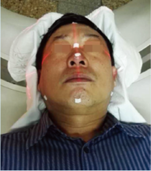
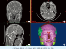
Objective To explore an efficient method for the establishment of three-dimensional (3-D) digital models of maxillofacial region including muscle tissue based on CT and MRI images fusion on a personal computer, integration of CT and MRI data, and provide accurate 3D model for biomechanical analysis.Methods A male volunteer was scanned on maxillofacial region by spiral CT and MRI. Two kinds of data obtained were imported into Mimics 15. In the three sections, namely the transverse, sagittal, coronal sections, two kinds of data were adjusted to the same anatomical layers. The most obvious anatomical points on each layer were selected as registration points. Then, the multi-points registration was implemented for data fusion. Then the bone and facial skin were segmented and 3D reconstructed using CT data, the main facial muscles were segmented and 3D reconstructed using MRI data.Results The 3D model including 3 pairs of masticatory muscles, 12 pairs of facial expression muscles, facial skin and jaw tissues were established.Conclusion The efficient registration and fusion of CT and MRI datas were accomplished. Moreover, this method can be used for further segmentation and reconstruction of other important structures in craniofacial area, such skin, blood vessel, fat, lymph node and the brain tissues.
Keyword:
Computed tomography; Magnetic resonance imaging; Maxillofacial region; Registration; Digital model
64排螺旋CT(Philips/Brilliance 64, Netherlands); MRI(GE, USA)。个人PC系统配置:Intel(R)Core(M)i7-6500CPU、3.20 GHz处理器、6 G DDR2内存、256 M RADEON显卡、4 TB硬盘, Wind7-SP1操作系统, Mimics15.0 (Materialise’ s Interactive Medical Image Control System, Belgium)。
KatsumuraS, SatoK, IkawaT, et al. “High-precision, reconstructed 3D model” of skull scanned by conebeam CT: Reproducibility verified using CAD/CAM data[J]. Leg Med (Tokyo), 2016, 18: 37-43. [本文引用:1]
KitamotoE, ChikuiT, KawanoS, et al. The application of dynamic Contrast-Enhanced MRI and Diffusion-Weighted MRI in patients with maxillofacial tumors[J]. Acad Radiol, 2015, 22(2): 210-216. [本文引用:1]
[6]
KoulisTA, DollCM, BrownD, et al. Implementation and validation of a combined MRI-CT-based cervical cancer brachytherapy program using existing infrastructure[J]. Brachytherapy, 2016, 15(3): 319-326. [本文引用:1]
[7]
WangY, LiuZY, DouWC, et al. Application of preoperative CT/MRI image fusion in target positioning for deep brain stimulation[J]. Chin Med Sci J, 2016, 31(3): 161-167. [本文引用:1]
HlubekRJ, TheodoreN, Chang SW. CT/MRI fusion for vascular mapping and navigated resection of a paraspinal tumor[J]. World Neurosurg, 2016, 89: 732. e7-732. e12. [本文引用:1]
[10]
LiuX, MeiW, DuH. Structure tensor and nonsubsampled shearlet transform based algorithm for CT and MRI image fusion[J]. Neurocomput, 2017, 235: 131-139. [本文引用:1]
LiP, LiZ, TianW, et al. A strategy for removal of foreign body in mand ible with navigation system[J]. Int J Oral Maxillofac Surg, 2015, 44(7): 885-888. [本文引用:1]
Objective To reconstruct three-dimensional (3D) image of posterior cruciate ligament (PCL) based on MRI and CT image fusion. Methods CT and MRI scans were performed on 12 knees of young men. The Dicom data were extracted and unified. The outline of PCL on MRI imaging was drew and plugged into the CT data. Finally, the visible 3D image of PCL with adjacent bones was reconstructed. The imaging anatomical measurements were examined and compared with those in published literature. Results Two cases were excluded from this study because of data deviations. The 3D visible reconstruction of PCL was proved to be feasible on the other ten cases. Conclusion Three-dimensional visible reconstruction of PCL based on CT and MRI image fusion is feasible, which can provide support for individualized treatment of PCL injuries. Further simplification with increased accuracy may be needed.
 , 艾毅龙
, 艾毅龙


